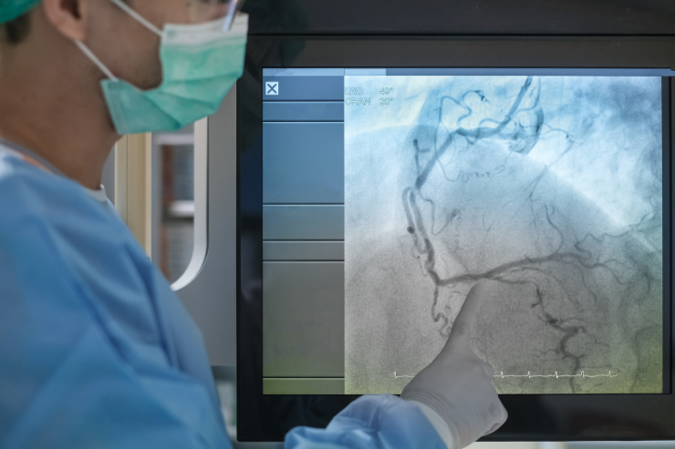
Angiogram

An angiogram is a diagnostic imaging procedure used to visualize the inside of blood vessels and organs. It employs X-ray technology combined with a contrast dye to produce detailed images of the blood vessels, helping to identify abnormalities such as blockages, narrowing, or malformations.
How Does an Angiogram Work?
During the procedure, a catheter is inserted into a blood vessel, usually through the groin or arm. This catheter is guided to the area of interest under X-ray guidance. Once positioned correctly, contrast dye is injected through the catheter into the blood vessels. The dye makes the vessels visible on X-ray images. These images are then analyzed by medical professionals to assess the condition of the blood vessels and detect any issues.
Types of Angiograms
- Coronary Angiogram: Focuses on the heart’s arteries to detect coronary artery disease.
- Cerebral Angiogram: Examines blood vessels in the brain to diagnose aneurysms or strokes.
- Pulmonary Angiogram: Used to identify clots or blockages in the lungs’ blood vessels.
- Peripheral Angiogram: Assesses blood flow in the limbs to diagnose conditions like peripheral artery disease.
Why is an Angiogram Important?
Angiograms are crucial for diagnosing and evaluating the severity of vascular conditions. They help guide treatment decisions, such as the need for angioplasty, stenting, or surgery. By providing detailed images of blood vessels, angiograms allow for timely and accurate medical interventions, improving patient outcomes and overall cardiovascular health.
